Compound vs stereo microscope
Compound for a thin sample that light can pass through. Stereo for a whole object you still need to turn by hand. The larger magnification number is not the better fit.
A compound microscope and a stereo microscope are built for different objects. The compound head sends light up through a thin specimen on a slide and can reach about 1000×. The stereo head looks down at a whole object, keeps a gap large enough for your fingers, and usually stays under 50×. Pick from the sample. The magnification printed on the box comes second.
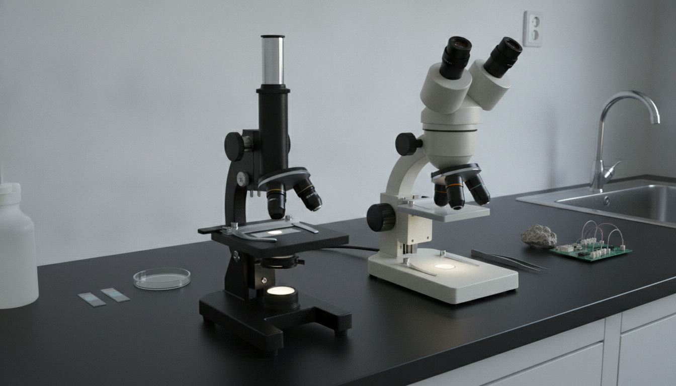
| Compound microscope | Stereo microscope | |
|---|---|---|
| Magnification | About 40×–1000× | About 7×–45× on a common zoom body |
| Working distance | A few millimeters at high power | Tens of millimeters |
| What you see | A flat slice | Depth, from two optical paths |
| Sample | Thin enough for light to pass through | Whole object, usually opaque |
| Light | From below, through the specimen | Onto the surface |
| Hands on the object | No room at high power | Room for fingers or a probe |
| Typical work | Cells, bacteria, thin tissue | Coins, insects, boards, solder joints |
What actually separates them
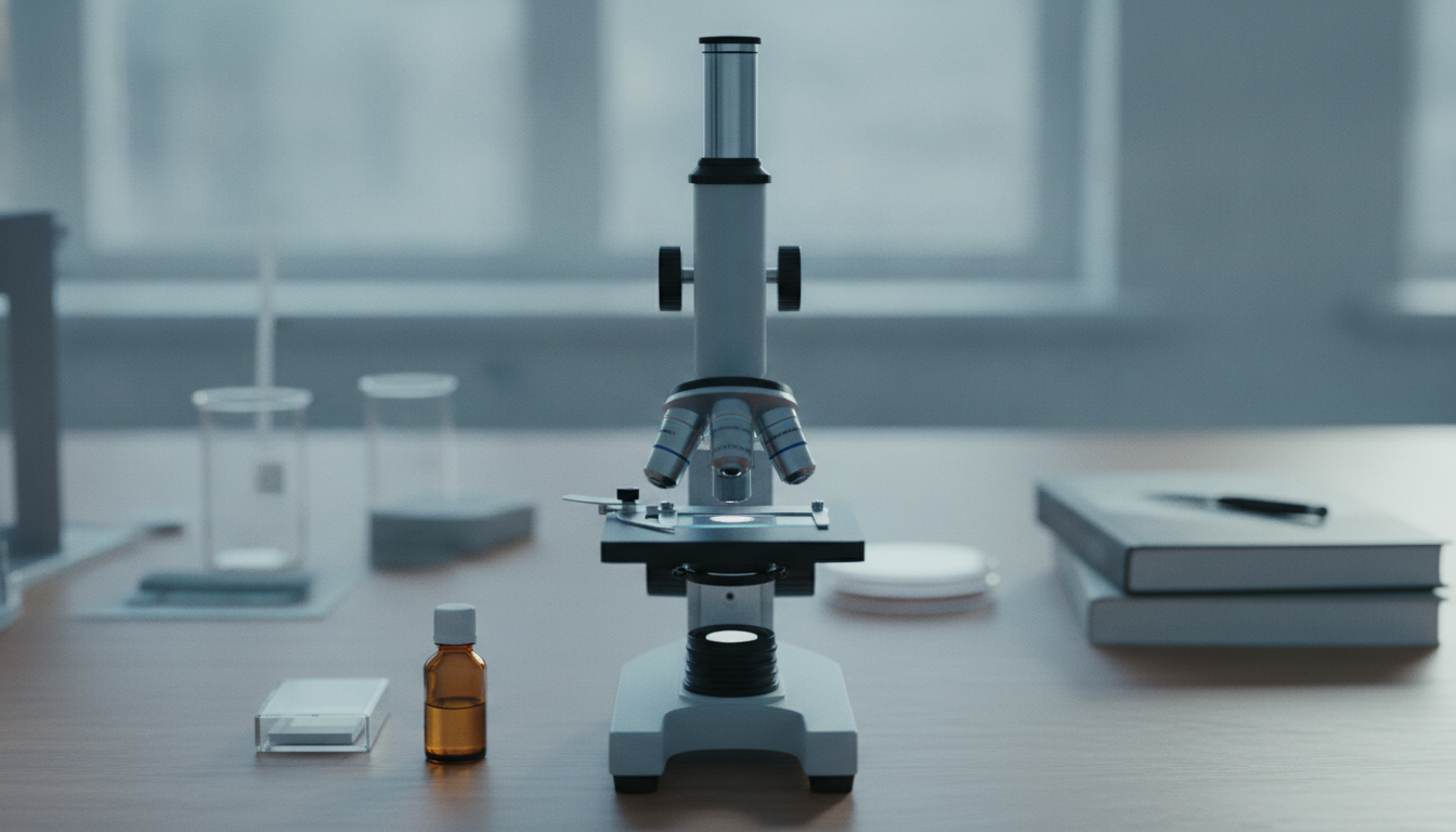
Magnification and working distance move in opposite directions on a compound stand. A 4× objective leaves a little room. A 40× objective is already close. A 100× objective sits so near the coverslip that a fingertip will not fit, and the lens often needs a drop of immersion oil between it and the glass. That closeness is what lets the lens collect enough light to resolve a cell. It is also why a coin, an insect, or a circuit board cannot go on that stand: the object is too thick, and the lens would hit it before the image is sharp.
A stereo body keeps that gap on purpose. Even at the top of a 0.7×–4.5× zoom there is usually room to turn a coin, move a specimen pin, or touch a solder joint while it stays in focus. The image has depth because each eye gets its own light path. A compound image is one flat plane. Anything above or below that plane goes soft.
The light follows the same split. A compound stand shines up through the slide. A condenser and an iris under the stage control how much of that light, and how wide a cone, reaches the specimen. If the specimen is too thick to pass light, the field stays dark no matter how strong the lamp is. A stereo stand lights the face of the object. A ring light flattens shadows on a board. Two gooseneck lamps, aimed from the sides, show the relief of a mint mark or a solder fillet.
How the magnification number is built
Total magnification is the eyepiece multiplied by the objective. A “20× eyepiece” line on its own does not say what you can resolve.
On a typical compound head the eyepieces are 10× and the objectives are 4×, 10×, 40×, and 100×. That is 40×, 100×, 400×, and 1000×. The 4× and 10× objectives are for finding the specimen and seeing how a tissue is arranged. The 40× objective is where cell detail starts. The 100× objective is the oil lens: without oil the image is dim and soft, and with oil the working distance is a fraction of a millimeter. Two 40× objectives with different numerical aperture do not show the same detail. NA, not the × mark, is the resolution number on a compound head.
On a common stereo zoom the body is marked 0.7×–4.5× and the eyepieces are 10×. The range you actually get is about 7×–45×. At 7× a whole coin fits in the field. At 45× you are looking at a mint mark or a single component, and you still have room to move the object. Some stereo heads skip the zoom and use a turret of fixed steps, often around 1×, 2×, and 4×, or 2× and 4×. A zoom is easier when you stay on one object and want to move in and out. Fixed steps are easier when you want the same magnification every time you come back to that lens.
Field of view shrinks as magnification rises. A stereo scope near 10× can hold a coin. The lowest step on a compound head is usually 40×, and even that shows a patch of a slide rather than the slide as an object. At 400× the field is a small disc of cells. Chasing a higher × number without checking the working distance and the field is how a stand ends up wrong for the object.
Match the sample
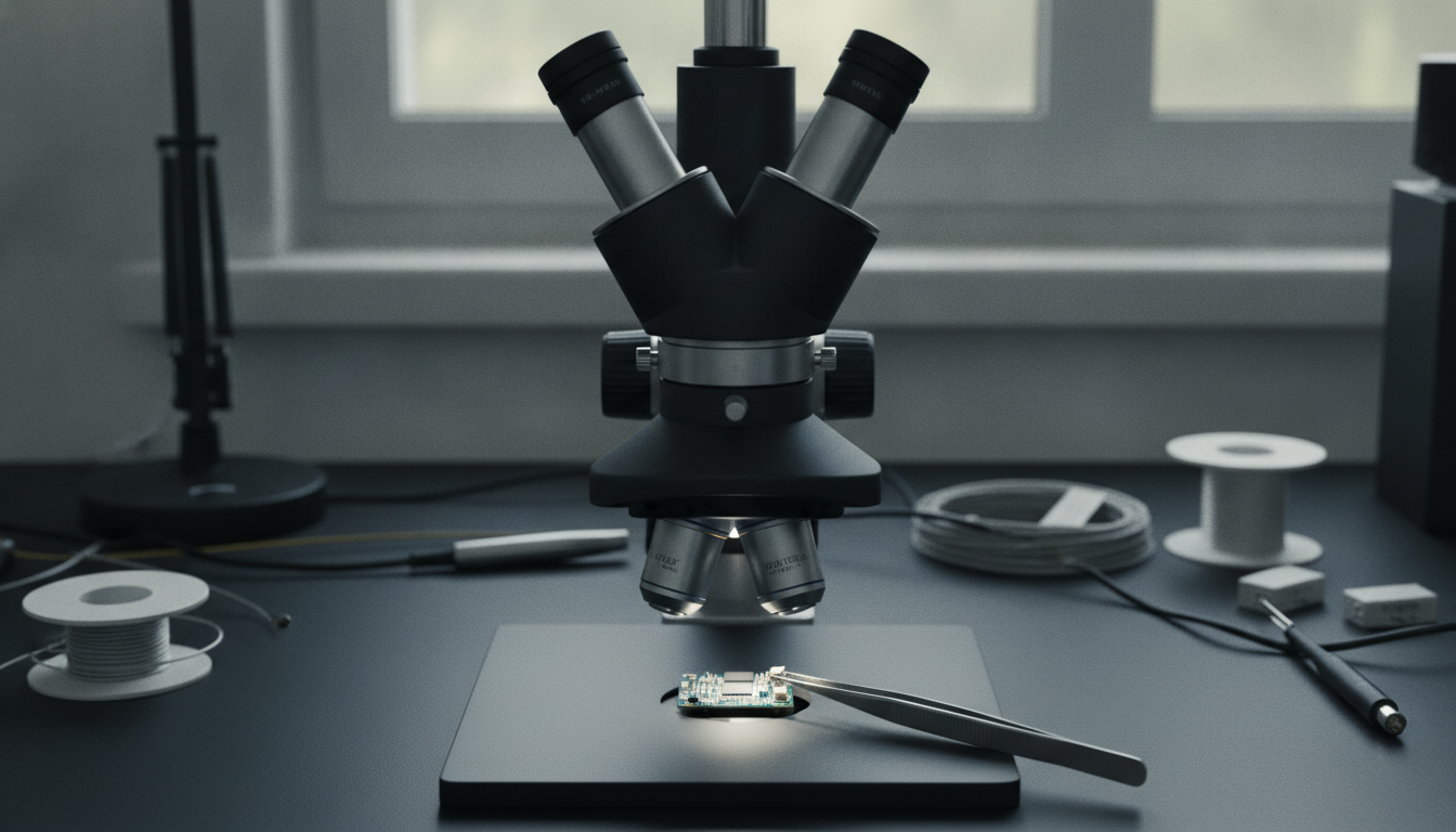
Hold the object before you choose. If it can be cut or smeared thin enough to pass light, and you need to see what is inside that thin layer, use a compound microscope. If it has to stay whole, and you need to see its surface in depth or move it while you look, use a stereo microscope.
- Cells, bacteria, blood smears, and thin tissue go on a compound stand. The specimen is already on a slide, or it can be.
- Insects, minerals, coins, circuit boards, and solder joints go on a stereo stand. Sectioning them would destroy the thing you came to inspect.
- A polished metal or rock slice already mounted on a slide goes on a compound stand. The sample is flat and thin, even though the original rock was not.
A camera on the head does not change this. A digital microscope is a camera on one of these two optical designs, or a simple USB camera with its own lens. The sensor does not decide whether light comes through the sample or off its surface. Read the working distance and the magnification range, then treat it as compound or stereo.
When you need both
One head does not cover the other. A biology student who looks at prepared slides and also dissects a flower or an insect needs both: the compound head for the slide, the stereo head for the whole specimen. A bench that repairs boards and also checks a soldered joint in cross-section has the same split. The cross-section, if it is thin and lit from below, is compound work. The populated board is stereo work.
If every object you care about is a slide, a stereo head will feel vague and under-powered. If every object is a coin, a board, or an insect you want to turn, a compound head will not focus on it at all. Adding the second head later is reasonable. Pretending one range of magnification covers both jobs is not.
Stand and light decide whether it is usable
The head gets the attention. The stand decides whether you can work. A short pillar is enough for slides: the specimen is small and it does not move. A boom arm clears the bench for a board or a large specimen, and it lets you swing the head out of the way. The same stereo head on a short pillar will not cover a circuit board. On a boom it will.
For surface work, a ring light is the even wash, and two gooseneck lamps are how you rake light across relief. On a compound stand the condenser and the iris already shape the light, so extra lamps are rare. A trinocular port, or a tube for a camera, matters only when you need a photo or a second person watching a screen. It does not change which objects the optics can take.
A wider zoom on a loose, wobbling pillar is worse than a shorter zoom on a steady base. The extra magnification is useless if the image jitters when you touch the focus knob.
Questions
Can a stereo microscope show cells or bacteria?
No. A typical stereo range tops out around 45×, sometimes near 50×, and the light is bouncing off a surface. Cells and bacteria need light coming through a thin specimen and a compound objective in the 40×–100× range, which is 400×–1000× at the eye.
Can a compound microscope inspect a coin or a circuit board?
No. The working distance at useful power is a few millimeters or less, and the lighting is meant to pass through a slide. A coin or a board never reaches focus as a whole object. Use a stereo head, with enough gap under the lens to turn the piece.
What does 1000× mean on a compound microscope?
It is almost always a 10× eyepiece times a 100× objective. That objective is meant to be used with immersion oil, and the front lens is nearly touching the coverslip. The number on the box is real only when both parts are in the light path and the oil is there. A 20× eyepiece on a low objective is not the same thing.
When do you need both?
When the work includes both slides and whole objects. Prepared tissue and a dissection, or a thin cross-section and a populated board, are two jobs. One magnification range does not cover both.
Is a digital microscope a third kind?
No. It is a camera. The optical head underneath is still compound or stereo, unless it is a simple close-up camera with no microscope optics at all. Judge it by working distance, whether light passes through the sample, and the real magnification after the eyepiece or the zoom, not by the word digital.